Home Research Lab Members Publications Collaborations Support Upcoming Events Contact Us
 
Research Interests
 
Spermatogonial proliferation in primates
 
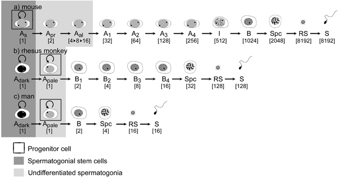
The diagram illustrates a comparison of germ cell expansion schemes in mouse, monkey and human testes. It is important to note that the primate testes contains in addition to a true stem cells (Ad) a population of progenitor cells (Ap) which undergo cyclic self-renewal and generation of progeny. The mouse testis does not have a progenitor type germ cell but a much more efficient strategy for mitotic expansion of germ cells after division of the stem cell (As).
 

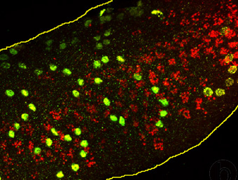
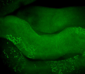
We developed a new strategy to detect BrdU in whole mounts of seminiferous tubules by immunofluorescence. The immunopositive cells can be seen as chains or cohorts of spermatogonia or preleptotene spermatocytes. Our data reveal that premeiotic germ cell expansion follows a clonal scheme.
 

wholemountb

wholemounta

When the BrdU localization (green) is combined with determination of acrosome development (acrosin staining, red) and confocal imaging, we are able to describe a stage-specific appearance of S-phase in rhesus monkey testes and to determine number and size of clones at each spermatogenic stage. We determined that spermatogenesis starts at stage VII with a first division of pairs or quadruplets of Ap spermatogonia. Clonal splitting after this initial division is followed by a second mitotic step after which no splitting occurs. Cells now arrest generating new Ap pairs or quadruplets or generate clonally expanding chains of B-spermatogonia.
 
 
Xenografting of primate testicular tissue
 
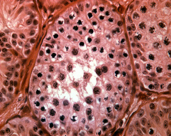
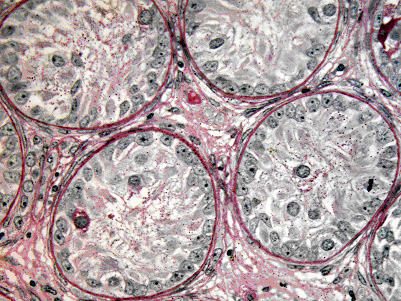
Xenografting of testicular tissue provides a strategy for preservation and differentiation of immature testicular tissue. The micrograph shows a crossection through seminiferous tubules from a rhesus monkey whose testes had been cryopreserved at the juvenile stage of development (only A-spermatogonia as germ cells). Three months after thawing and xenografting spermatogenesis is initiated up to the level of meiosis.
   
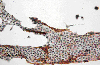
Xenografting of monkey tissue is a powerful tool to explore the effects of drugs or treatments on the developing monkey testis. After small fragments of juvenile rhesus monkey testes were grafted under the back skin of nude mice, spermatogenesis was initiated up to the level of spermatocytes after 3 months. The mice were then treated with busulfan or vehicle. Four weeks later, busulfan treated mice showed a depletion of B-spermatogonia and more advanced germ cells. The micrograph shows seminiferous tubules of a monkey testicular xenograft with Sertoli cells and A-spermatogonia as the most advanced germ cell types.
   
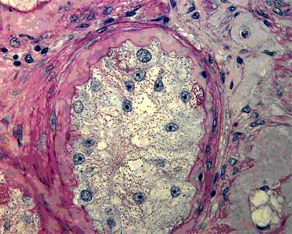
Human ectopic xenografting was performed using adult testes. Grafting of adult testicular tissue is generally less successful compared to the immature testis. The micrograph shows limited survival of seminiferous tubules in xenografts from an adult transsexual patient. The patient had been treated with high dose of estrogens leading to testicular atrophy prior to grafting of the tissue. The grafted tissue shows fibrosis in the tubular wall and limited survival of Sertoli cells and spermatogonia.
 
 
Spermatogonial isolation and transplantation


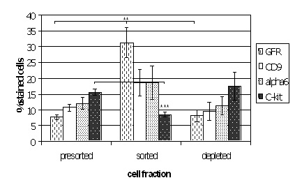
Magnetic activated cell sorting was applied to enrich spermatogonial stem cells from immature mouse testes. After isolation the cells were stained immunohistochemically for additional spermatogonial markers. The plot shows that the sorted fraction was significantly enriched for GFRA-1 when compared to presorted and depleted fractions. In addition, a significant depletion of c-kit positive cells was encountered. These data show that the technique can be applied to obtain enriched fractions of spermatogonial stem cells.
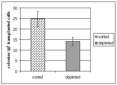
 

The enrichment of spermatogonial stem cells was confirmed by germ cell transplantation experiments performed in collaboration with Kyle Orwig, Pittsburgh Development Center, Magee Woman’s Research Institute. Mouse testes receiving sorted cell fractions generated significantly more spermatogenic colonies in busulfan depleted recipients when compared to mice receiving cells from the depleted fraction.
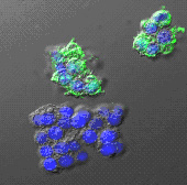
 
The micrograph depicts confocal images of isolated spermatogonial stem cells using antibodies directed against GFRA-1. After GFRA-1 based enrichment, the cells were seeded on slides and immunohistochemically stained for OCT-4 (green fluorescent labeling). Some of the isolated spermatogonia stain positive for this pluripotency marker while other are fully negative. These studies were performed in collaboration with Ahmi Ben-Yehudah (Pittsburgh Development Center, Magee Woman’s Research Institute).
   
   
Aging of spermatogonial stem cells
 

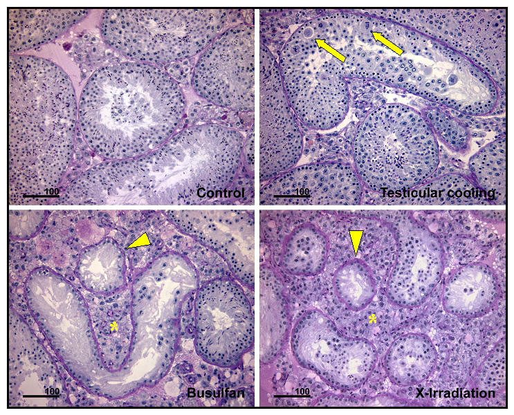
We explore the effects of aging on testicular stem cells in mouse and monkey models. This plate of micrographs shows the effect of cooling, busulfan and X-irradiation on the testis of old Balb-c mice. In comparison to control testes, minor disorganization is observed after cooling. In contrast, busulfan and X-irradiation show a severe depletion of spermatogenic cells. This study revealed that the degree of damage following these exposures are similar in old and young mice.
 

The two plots show the effect of testicular irradiation (4Gy) on individual testicular volume (left panel) and sperm counts (middle panel) in groups of young adult (red) and aging (blue) rhesus monkeys. Radiation exposure evoked an almost complete depletion of germ cells as becomes evident from a representative testicular biopsy (Mk3058) obtained 8 weeks after irradiation shown as micrograph in the right panel.
 
 
Testicular Morphogenesis
   
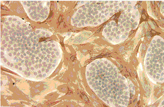
The micrograph shows mixed cultures of peritubular and Sertoli cells from immature rat testes. The cells undergo well defined morphogenetic changes. In this picture Sertoli cells form island like structures between whirls of peritubular cells.
   
This micrograph depicts the next stage of in vitro morphogenesis. Sertoli cells aggregate and proliferate to form cord like structures as the final stage of differentiation on laminin coated coverslips.
   
The same single cell suspensions as above can be cultured in matrigel forming three-dimensional aggregates of Sertoli cells. When xenografted under the back skin of nude mouse recipients these fragments of rat peritubular/Sertoli cells develop into the depicted seminiferous tubules. These studies reveal that Sertoli cell carry the full potential for morphogenesis of testicular tissue.
   
 
  Home Research Lab Members Publications Collaborations Support Upcoming Events Contact Us