 |
|
How our results mesh with
the crystal structure of the nucleosome |
|
 |
|
How our results mesh with
the crystal structure of the nucleosome |
|

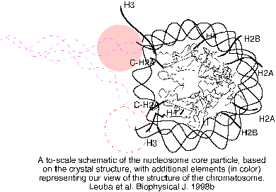
A to-scale schematic of the core particle, based on the crystal structure (Luger, K., A. W. Mader, R. K. Richmond, D. F. Sargent, and T. J. Richmond. 1997. Nature, 389, 250-260) with additional elements (globular domain GD of linker histone and adjacent stretches of linker DNA), representing our view of the structure of the chromatosome. The structure of the core particle is a sketched outline of figure 1A from Lugar et al. (1997), with line traces of the paths of the DNA gyres and the backbones of the histone molecules in the octamer. The beige circle outside of the particle is the globular domain of histone H5 drawn to scale and is situated off-axis, as proposed by Travers and Muyldermans (Travers, A. A. and S. V. Muyldermans. 1996. J. Mol. Biol. 257, 486-491), and more recently by us ( An, W., Leuba, S. H., van Holde, K., and Zlatanova, J. 1998. Proc. Natl. Acad. Sci. 95, 3396-3401). The symmetrically located dashed circle represents the alternative off-axis position of the GD. It should be noted that the exact location of the GD is not important for the present model; a central over-the-axis location is equally compatible with the proposed interactions. Linker DNA is arbitrarily drawn as dashed double helices going out of the core particle. Note the location of the tails of the core histones (labeled thick lines), particularly the proximity of those of histone H3 to linker DNA.